An international team led by the ITACA Institute of the Universitat Politècnica de València (UPV) has developed one of the most complete and detailed structural atlases of the human brain to date. Called HoliAtlas, it will be especially useful for the study and early diagnosis of neurological and neurodegenerative diseases such as Alzheimer’s or Parkinson’s.
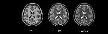
The new map is based on ultra-high-resolution multimodal magnetic resonance imaging (MRI) and far exceeds the level of detail of existing MRI-based atlases.
The work, published in the journal Scientific Reports (Nature), has been led by Professor José V. Manjón, coordinator of the MIALAB group at ITACA-UPV, in collaboration with international institutions such as the CNRS and the University of Bordeaux, as well as Spanish and European centers.
More personalized diagnoses and treatments
HoliAtlas offers a complete, multilevel representation of the brain, from global structures to very specific substructures, in a holistic way. Its resolution and multimodal integration facilitate the identification of deep structures and enable the development of more precise automatic segmentation methods, improve morphological analysis, and detect very subtle anatomical changes.
“For this reason, this atlas could be of great help in studying pathologies such as Alzheimer’s or Parkinson’s and enable more accurate diagnoses. Having increasingly precise brain atlases is key to understanding the architecture of the human brain, integrating data from different studies, and advancing toward more personalized diagnoses and treatments”, explains José Vicente Manjón, head of the MIA-LAB group at ITACA-UPV.
A leap in resolution and anatomical detail
Brain atlases function as reference maps, essential for accurately locating anatomical structures and comparing data across studies or populations. They are also key tools in both neuroscience research and clinical applications, such as surgical planning and the analysis of neurological diseases.
“Until now, most MRI-based atlases had an approximate resolution of 1 mm³. The new atlas reaches a resolution of 0.125 mm³, allowing observation of much smaller and more complex brain structures”, notes Sergio Morell, researcher in the MIA-LAB group at ITACA and co-author of the study.
Based on data from 75 healthy brains
To build this atlas, the researchers used brain images from 75 healthy volunteers from the prestigious Human Connectome Project, one of the largest international repositories of neuroimaging data. From these data, they applied advanced processing and normalization techniques to generate an average brain model.
“At its most detailed level, the atlas includes up to 350 anatomical regions obtained through the integration of seven different segmentation protocols, combining neuroanatomical analysis tools, artificial intelligence algorithms, and expert manual corrections,” concludes Sergio Morell.
REFERENCE: José V. Manjón, Sergio Morell-Ortega, Marina Ruiz-Perez, Boris Mansencal, Edern Le Bot, Marien Gadea, Enrique Lanuza, Gwenaelle Catheline, Thomas Tourdias, Vincent Planche, Remi Giraud, Denis Rivière, Jean-Francois Mangin, Nicole Labra-Avila, Roberto Vivo-Hernando, Gregorio Rubio, Fernando Aparici-Robles, Maria de la Iglesia-Vaya & Pierrick Coupé. Ultra-high resolution multimodal MRI densely labelled holistic structural brain atlas. Scientific Reports. NATURE. DOI: https://www.nature.com/articles/s41598-026-40186-2